The Transmission Electron Microscope (TEM), a type of electron microscope, is one of the most powerful instruments in contemporary biological and materials research. It has profoundly enhanced our understanding of cellular organization and biomolecular architecture by providing images at resolutions far beyond those achievable with light microscopes. Unlike optical microscopes that employ visible light and glass lenses, the TEM utilizes a focused beam of electrons and electromagnetic lenses to generate highly magnified images of thin specimens. For students and researchers in biology, the TEM serves as a fundamental tool for exploring the ultrastructure of cells, tissues, and macromolecular complexes.
Principle of Transmission Electron Microscope
The Transmission Electron Microscope operates on the principle of electron–matter interaction. According to the de Broglie hypothesis, electrons behave as waves, and their wavelength is inversely proportional to their momentum. Because the wavelength of accelerated electrons is several orders of magnitude shorter than that of visible light, the TEM can resolve much finer structural details — even approaching the atomic scale [1].
J Brew, uploaded on the English-speaking Wikipedia by en:User:Hat’nCoat., CC BY-SA 3.0, via Wikimedia Commons
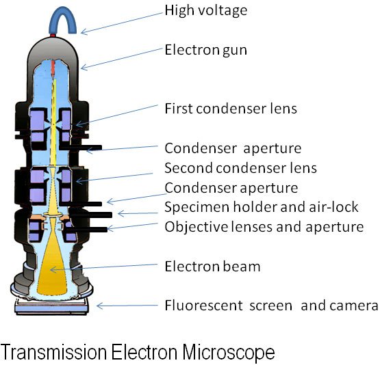
In TEM operation, electrons are emitted from an electron gun (commonly a tungsten filament or a field emission source) and accelerated through a potential difference typically ranging between 60 kV and 300 kV. The accelerated electron beam passes through a vacuum column and is focused onto a specimen using electromagnetic lenses, which serve a function analogous to optical lenses in light microscopy. As the beam interacts with the specimen, some electrons are transmitted, while others are scattered or absorbed, depending on the specimen’s density and composition. The transmitted electrons are focused by objective and projector lenses to form an image on a fluorescent screen or digital camera sensor [2].
| You may also like NOTES in... | ||
|---|---|---|
| BOTANY | BIOCHEMISTRY | MOL. BIOLOGY |
| ZOOLOGY | MICROBIOLOGY | BIOSTATISTICS |
| ECOLOGY | IMMUNOLOGY | BIOTECHNOLOGY |
| GENETICS | EMBRYOLOGY | PHYSIOLOGY |
| EVOLUTION | BIOPHYSICS | BIOINFORMATICS |
Dr Graham Beards, Public domain, via Wikimedia Commons
Key aspects of the TEM principle:
- Electron Source: Produces a coherent, high-energy beam of electrons.
- Electromagnetic Lenses: Replace glass lenses used in optical microscopes.
- Specimen Thinness: Samples must be extremely thin (typically 50–100 nm) to permit electron transmission.
- Image Formation: Based on differential electron scattering and transmission that produce contrast.
The final image is a projection in which dense regions (that scatter more electrons) appear dark, while less dense regions appear lighter.
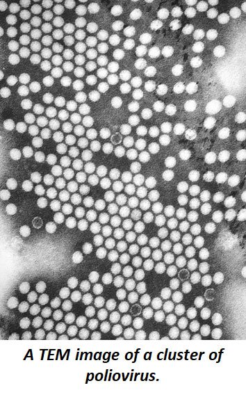
Photo Credit:Content Providers(s): CDC/ Dr. Fred Murphy, Sylvia Whitfield, Public domain, via Wikimedia Commons
Sample Preparation for TEM
Since electrons cannot penetrate thick or hydrated materials, biological specimens must undergo extensive preparation to make them suitable for observation in a TEM. The standard preparation involves the following stages [3]:
- Fixation: The first step stabilizes and preserves cellular structures. Glutaraldehyde cross-links proteins, while osmium tetroxide stabilizes lipids and enhances contrast.
- Dehydration: Water is gradually removed using graded ethanol or acetone series, as water is incompatible with the vacuum environment of the TEM.
- Embedding: The specimen is infiltrated with a resin such as epoxy or acrylic, which hardens and provides mechanical stability.
- Sectioning: Ultrathin sections (approximately 50–100 nm thick) are cut using an ultramicrotome fitted with a diamond or glass knife.
- Staining: To enhance electron scattering and improve contrast, sections are stained with heavy metals such as lead citrate and uranyl acetate.
Proper preparation is essential because poorly fixed or stained samples can lead to image artifacts that misrepresent the actual cellular architecture.
Applications of the Transmission Electron Microscope in Biology
The TEM has a broad range of applications in biological and biomedical sciences due to its ability to visualize subcellular and macromolecular structures in great detail:
- Cell Biology: TEM reveals the ultrastructure of organelles such as mitochondria, ribosomes, nuclei, endoplasmic reticulum, and Golgi apparatus, enabling detailed studies of cell organization and function [4].
- Virology: Viruses that are too small to be resolved with light microscopes can be visualized and characterized using TEM. It provides valuable information about viral morphology, size, and assembly processes [5].
- Microbiology: TEM assists in studying bacterial and archaeal morphology, including cell walls, flagella, and pili, contributing to taxonomy and microbial physiology research.
- Pathology: In medical diagnostics and pathology, TEM detects ultrastructural changes associated with diseases, including mitochondrial abnormalities, inclusion bodies, and disruptions in membrane integrity [6].
- Molecular Biology and Structural Biochemistry: TEM allows visualization of macromolecular complexes, such as ribosomes, cytoskeletal filaments, and protein assemblies. Modern techniques such as cryo-electron microscopy (cryo-TEM) have further advanced the field, allowing structures to be studied in their near-native hydrated states without staining or embedding [7].
Advantages of the Transmission Electron Microscope
- Exceptional Resolution: The TEM can achieve resolutions up to 0.1–0.2 nanometers, far surpassing the diffraction limit of light microscopy.
- Detailed Structural Visualization: Enables high-resolution imaging of organelles, viruses, and macromolecules.
- Analytical Versatility: When combined with techniques such as energy-dispersive X-ray spectroscopy (EDS), the TEM can provide both structural and elemental composition data.
Limitations of the Transmission Electron Microscope
Despite its remarkable capabilities, the TEM is not without drawbacks:
- Complex and Time-Consuming Sample Preparation: Requires specialized equipment and expertise.
- Vacuum Environment: Samples must be dehydrated and cannot be observed in their living state.
- High Cost and Maintenance: TEM instruments are expensive and demand trained personnel and stable infrastructure.
Conclusion
The Transmission Electron Microscope stands as a cornerstone of modern biological and material science research. By exploiting the wave nature of electrons, it reveals the minute details of cellular and molecular architecture that are invisible to light microscopy. Although the technique requires intricate specimen preparation and specialized training, its unparalleled resolution and versatility make it indispensable for understanding the organization and function of biological systems. For students and researchers alike, mastering the principles and applications of the TEM is crucial for advancing in fields such as cell biology, virology, microbiology, and molecular structural analysis.
Download PDF – Transmission Electron Microscope
References
- Williams, D. B., & Carter, C. B. (2009). Transmission Electron Microscopy: A Textbook for Materials Science (2nd ed.). Springer.
- Reimer, L., & Kohl, H. (2008). Transmission Electron Microscopy: Physics of Image Formation and Microanalysis (5th ed.). Springer.
- Bozzola, J. J., & Russell, L. D. (1999). Electron Microscopy: Principles and Techniques for Biologists (2nd ed.). Jones and Bartlett Publishers.
- Alberts, B., Johnson, A., Lewis, J., et al. (2015). Molecular Biology of the Cell (6th ed.). Garland Science.
- Noda, T., & Kawaoka, Y. (2010). Structure of influenza virus and its implications for virus assembly and budding. Future Microbiology, 5(4), 523–531.
- Walker, R. A. (2004). Electron microscopy in diagnostic pathology. Histopathology, 44(3), 191–197.
- Kühlbrandt, W. (2014). The resolution revolution in cryo-EM. Nature Structural & Molecular Biology, 21(9), 819–823.
FAQ on Transmission Electron Microscope
Short Answer Questions (Answer in 1–2 sentences.)
- Who developed the Transmission Electron Microscope? (Remember)
- What is the main principle behind the Transmission Electron Microscope? (Understand)
- Why is a vacuum required in a TEM? (Understand)
- What is the typical accelerating voltage used in a TEM? (Remember)
- Name two commonly used fixatives in TEM specimen preparation. (Remember)
- What is the typical thickness of a specimen section for TEM analysis? (Remember)
- How is contrast generated in a TEM image? (Understand)
- What type of lenses are used in a TEM instead of glass lenses? (Remember)
- Mention one major advantage and one limitation of the TEM. (Understand)
- Define the term “resolution” in the context of TEM. (Remember)
Short Essay Questions (Answer in 4–6 sentences or one short paragraph.)
- Explain how the de Broglie hypothesis is relevant to the principle of the Transmission Electron Microscope. (Understand)
- Describe the major components of a TEM and state the function of each. (Understand)
- Discuss the importance of thin sectioning in TEM sample preparation. (Apply)
- Outline the sequential steps in the preparation of a biological specimen for TEM observation. (Remember/Understand)
- Compare the working principles of the Transmission Electron Microscope (TEM) and the Scanning Electron Microscope (SEM). (Analyze)
- Explain how heavy metal staining enhances image contrast in TEM. (Apply)
- Describe how TEM can be applied to study viruses and their structures. (Apply)
- Explain the role of electromagnetic lenses in focusing the electron beam. (Understand)
- Analyze the advantages and disadvantages of using TEM for biological research. (Analyze)
- Discuss how cryo-electron microscopy has improved upon the limitations of conventional TEM. (Evaluate)
Essay Questions (Answer in about 300–400 words.)
- Describe in detail the working principle of the Transmission Electron Microscope.
Include the electron source, acceleration system, electromagnetic lenses, and image formation process. (Understand/Analyze) - Evaluate the role of the Transmission Electron Microscope in modern biological sciences.
Discuss its applications in cell biology, virology, microbiology, and pathology, highlighting both advantages and limitations. (Evaluate) - Propose modern improvements or techniques that can minimize artifacts in TEM specimen preparation.
Discuss challenges such as fixation, dehydration, and sectioning, and include innovations like cryo-TEM and vitrification. (Create)
Download Answer Key of these Questions PDF
<<< Back to Biophysics Quick Notes Page
🌱 Thanks for checking out this post on Principle of Transmission Electron Microscope
I’d love to know what you think—did you enjoy it, have any questions, or maybe a fun fact to add? 🤔✨
Drop your thoughts in the comments—I read them all and they really keep me motivated to share more cool Biology stuff with you! 💬💚
-Admin, EasyBiologyClass
| You may also like... | ||
|---|---|---|
| NOTES | QUESTION BANK | COMPETITIVE EXAMS. |
| PPTs | UNIVERSITY EXAMS | DIFFERENCE BETWEEN.. |
| MCQs | PLUS ONE BIOLOGY | NEWS & JOBS |
| MOCK TESTS | PLUS TWO BIOLOGY | PRACTICAL |