The Scanning Electron Microscope (SEM), a type of electron microscope, is a powerful analytical tool that revolutionized imaging in biological sciences. Unlike conventional light microscopes that rely on visible light, the SEM uses a concentrated beam of electrons to generate high-resolution, three-dimensional images of specimen surfaces. For biology students and researchers, the scanning electron microscope offers a deeper understanding of biological form and function, revealing intricate details that are invisible under optical microscopes. This blog post explains the Principle of Scanning Electron Microscope and its Applications.
Principle of Scanning Electron Microscope
The working principle of the scanning electron microscope is based on the interaction between electrons and the atoms of a specimen’s surface. An electron gun produces a fine beam of electrons, which is accelerated under high voltage (usually between 1–30 kV) and directed toward the specimen. The beam is focused and controlled by a series of electromagnetic lenses and scanning coils, allowing it to move across the surface in a raster (grid-like) pattern.
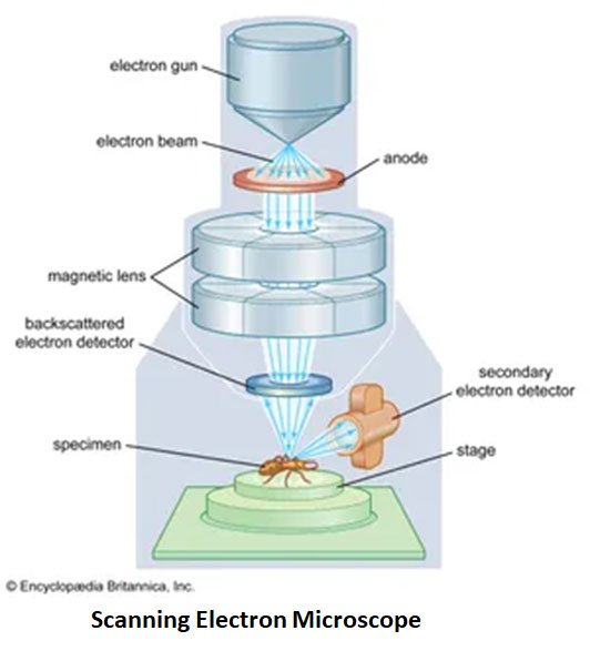
https://www.britannica.com/technology/scanning-electron-microscope#/media/1/526571/110970
When the primary electrons strike the specimen, they interact with its surface atoms, producing various types of signals, including secondary electrons, backscattered electrons, and X-rays. Among these, secondary electrons are most commonly used for imaging because they provide detailed information about the surface topography of the sample. These emitted signals are collected by detectors, converted into electrical signals, and displayed as a high-resolution image on a monitor. The resulting image represents the three-dimensional structure and texture of the specimen’s surface.
Key Components and Processes in SEM Operation:
- Electron Beam: Generated by a tungsten filament or field emission gun and accelerated to high energy.
- Scanning System: Moves the focused beam systematically across the specimen’s surface.
- Signal Detection: Detects secondary electrons for topographical imaging and backscattered electrons for compositional contrast.
- Image Formation: The signals are amplified, processed, and displayed to create a detailed 3D image.
| You may also like NOTES in... | ||
|---|---|---|
| BOTANY | BIOCHEMISTRY | MOL. BIOLOGY |
| ZOOLOGY | MICROBIOLOGY | BIOSTATISTICS |
| ECOLOGY | IMMUNOLOGY | BIOTECHNOLOGY |
| GENETICS | EMBRYOLOGY | PHYSIOLOGY |
| EVOLUTION | BIOPHYSICS | BIOINFORMATICS |
Sample Preparation for Scanning Electron Microscopy
Biological samples require careful preparation to endure the SEM’s high-vacuum environment and prevent damage from the electron beam. The preparation steps typically include:
- Fixation: The specimen is treated with fixatives such as glutaraldehyde to preserve cellular structures and prevent degradation.
- Dehydration: Water is gradually removed using a graded ethanol or acetone series, as moisture can distort the specimen in a vacuum.
- Drying: Critical point drying replaces the remaining liquid with gas, avoiding collapse of delicate structures.
- Coating: A thin conductive layer, usually gold, platinum, or carbon, is sputter-coated on the sample to prevent charging and improve image clarity.
Applications of the Scanning Electron Microscope in Biology
The SEM has a wide range of applications across biological disciplines:
- Cell Biology: Enables visualization of cell surfaces, microvilli, cilia, and cell-to-cell interactions.
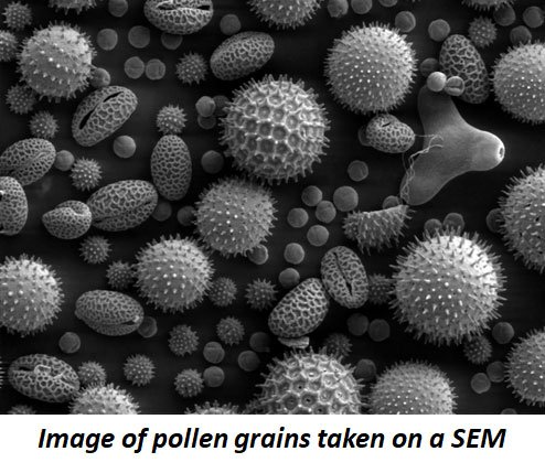
- Botany: Reveals detailed structures of leaf surfaces, trichomes, pollen grains, and stomatal complexes.
- Zoology: Used to study insect exoskeletons, scales, feathers, and other surface adaptations.
- Microbiology: Provides high-resolution images of bacterial colonies, spores, and biofilms.
- Pathology: Assists in examining tissue surface alterations and microstructural changes in diseased states.
Advantages of Scanning Electron Microscopy
- Produces detailed, high-resolution 3D images of specimen surfaces.
- Offers a large depth of field for viewing extensive surface areas in sharp focus.
- Suitable for studying a wide variety of biological materials, from microorganisms to complex tissues.
Limitations of Scanning Electron Microscopy
- Only surface features can be visualized; internal structures require transmission electron microscopy (TEM).
- Operation in vacuum conditions prevents observation of living specimens.
- Sample preparation is time-consuming and may introduce minor structural artifacts.
- Equipment is expensive and requires skilled operation and maintenance.
Conclusion
The scanning electron microscope has become a fundamental tool in biological research and education. By providing precise, three-dimensional visualization of surface morphology, it deepens our understanding of cellular and tissue organization. Although restricted to surface studies and requiring meticulous sample preparation, the SEM continues to play a central role in exploring biological complexity. For modern biology students and researchers, mastering the principles and applications of the scanning electron microscope opens the door to a world of microscopic detail and discovery.
Download PDF: Quick Note on Scanning Electron Microscope
FAQ on Scanning Electron Microscope (SEM)
Short-Answer Questions: Answer each question briefly (1–2 sentences).
- What is the full form of SEM? (Remembering)
- Who invented the scanning electron microscope? (Remembering)
- What type of beam is used in SEM? (Remembering)
- Mention one main difference between light microscopy and SEM. (Understanding)
- What is the usual voltage range used to accelerate electrons in SEM? (Remembering)
- Name two types of signals produced when the electron beam strikes the specimen. (Understanding)
- What is the main function of electromagnetic lenses in SEM? (Understanding)
- Why is gold or platinum coating applied to biological samples before SEM observation? (Understanding)
- What is meant by “secondary electrons” in SEM imaging? (Understanding)
- State one advantage and one limitation of SEM. (Understanding)
Short-Essay Questions: Answer each in about 4–6 sentences.
- Explain how the interaction between electrons and specimen atoms results in image formation in SEM. (Applying)
- Describe the purpose of the vacuum system in a scanning electron microscope. (Applying)
- Outline the steps involved in biological specimen preparation for SEM analysis. (Applying)
- How does SEM provide three-dimensional images compared to optical microscopy? (Analyzing)
- Differentiate between secondary and backscattered electrons in terms of their origin and information provided. (Analyzing)
- Discuss how critical point drying preserves delicate biological structures for SEM observation. (Applying)
- Explain why SEM is particularly useful in microbiology and botany. (Applying)
- Describe how the scanning system controls the movement of the electron beam across the specimen surface. (Applying)
- Analyze the impact of improper sample preparation on the quality of SEM images. (Analyzing)
- Compare SEM with TEM in terms of the type of information each provides. (Analyzing)
Essay Questions: Answer in 300–500 words.
- Evaluate the role of the scanning electron microscope in advancing modern biological research. Discuss its advantages and limitations in detail with relevant examples. (Evaluating)
- Analyze the importance of proper sample preparation in SEM and suggest improvements or modern techniques that minimize specimen distortion in biological imaging. (Analyzing / Evaluating)
- Design an experimental study where SEM could be used to investigate a specific biological structure (e.g., pollen grains, insect exoskeletons, or bacterial biofilms). Describe the steps, expected observations, and significance of the findings. (Creating)
Download the Answer Key of These Questions (PDF)
<<< Back to Biophysics Quick Notes Page
🌱 Thanks for checking out this post on Principle of Scanning Electron Microscope
I’d love to know what you think—did you enjoy it, have any questions, or maybe a fun fact to add? 🤔✨
Drop your thoughts in the comments—I read them all and they really keep me motivated to share more cool Biology stuff with you! 💬💚
-Admin, EasyBiologyClass
| You may also like... | ||
|---|---|---|
| NOTES | QUESTION BANK | COMPETITIVE EXAMS. |
| PPTs | UNIVERSITY EXAMS | DIFFERENCE BETWEEN.. |
| MCQs | PLUS ONE BIOLOGY | NEWS & JOBS |
| MOCK TESTS | PLUS TWO BIOLOGY | PRACTICAL |