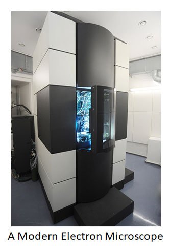
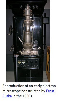
Why Electron Microscopy Matters?
The microscopic world extends beyond the reach of light microscopes, which are limited to resolving details down to about 0.2 µm, leaving structures like viruses (20–300 nm) and ribosomes (25–30 nm) invisible. Electron microscopy (EM) overcomes this barrier by using electron beams to achieve much higher resolution, revolutionizing biology and other sciences with detailed views of cellular ultrastructure, viruses, and even the surfaces of materials. This blog post introduces the principles of EM, its main types, specimen preparation methods, advantages and limitations, and its wide-ranging applications in biology, medicine, and materials science, highlighting its indispensable role in modern research.
Biophysics Quick Notes | Biophysics PPTs | Biophysics MCQs
This blog post explores the principles behind electron microscopes, the types of EM, specimen preparation, advantages and limitations, and their diverse applications in biology, medicine, and materials science. By the end, you’ll understand why EM is indispensable in modern scientific research.
Alice im Miniland, CC BY-SA 4.0, via Wikimedia Commons
How Electron Microscopes Differ from Light Microscopes?
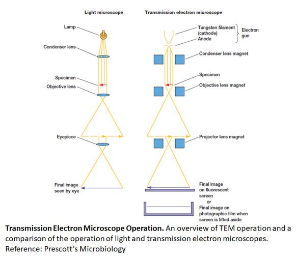
The most fundamental difference between electron and light microscopes is the nature of the illumination source. While light microscopes use photons to illuminate specimens, electron microscopes use a beam of high-energy electrons.
Why Use Electrons?
- Wavelength: The resolution of a microscope is limited by the wavelength of its illumination source. Visible light has a wavelength of approximately 400–700 nanometers, which restricts the ability to resolve structures smaller than 200 nanometers. Electrons, however, behave like waves with wavelengths in the sub-nanometer range at high velocities, enabling EM to resolve details at the atomic or near-atomic level.
- Electromagnetic Lenses: Unlike glass lenses used in light microscopy, electron microscopes use electromagnetic coils to focus the electron beam, bending electrons much like glass bends light.
- Vacuum Environment: Because electrons scatter in air, EM requires a high-vacuum chamber to maintain a clear beam.
| You may also like NOTES in... | ||
|---|---|---|
| BOTANY | BIOCHEMISTRY | MOL. BIOLOGY |
| ZOOLOGY | MICROBIOLOGY | BIOSTATISTICS |
| ECOLOGY | IMMUNOLOGY | BIOTECHNOLOGY |
| GENETICS | EMBRYOLOGY | PHYSIOLOGY |
| EVOLUTION | BIOPHYSICS | BIOINFORMATICS |
These features allow electron microscopes to achieve resolutions that light microscopes cannot, making them essential for examining the ultrastructure of cells, viruses, and even nanomaterials.
Principles of Electron Microscopy
Electron microscopy operates on a few core principles:
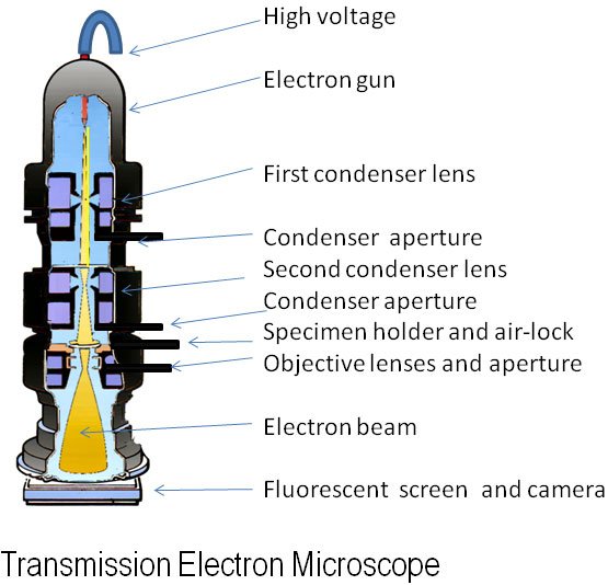
Electron Source (Electron Gun):
- Electrons are generated by an electron gun, often using a tungsten filament or a field emission source.
- A high-voltage potential accelerates the electrons to high velocities, shortening their wavelength and improving resolution.
Electromagnetic Lenses:
- Electron beams are focused using magnetic fields rather than glass lenses.
- Condenser lenses control beam intensity and focus, while objective lenses sharpen the final image.
Vacuum Environment:
- The microscope chamber is evacuated to prevent electrons from scattering in the air.
- This ensures that the electron beam remains coherent and accurately focused on the specimen.
Detectors and Imaging:
- Electrons interact with the specimen and are collected by detectors, which convert electron signals into images on a screen or computer.
- In transmission electron microscopy (TEM), electrons pass through the specimen; in scanning electron microscopy (SEM), electrons bounce off the surface.
The resolution of an electron microscope is proportional to the electron wavelength, which decreases as the electrons’ accelerating voltage increases. This is why high-voltage electron microscopes are capable of resolving structures down to fractions of a nanometer.
Dr Graham Beards, Public domain, via Wikimedia Commons
Types of Electron Microscopes
There are several types of electron microscopes, each optimized for specific applications.
Transmission Electron Microscope (TEM)
Principle: TEM works by passing a beam of electrons through an ultra-thin specimen. Denser regions scatter more electrons and appear darker in the final image, while less dense areas allow more electrons to pass and appear brighter.
Image Characteristics:
- Two-dimensional (2D) images of internal structures.
- Extremely high resolution—often <1 nm.
Applications:
- Visualizing organelles such as mitochondria, Golgi apparatus, or endoplasmic reticulum.
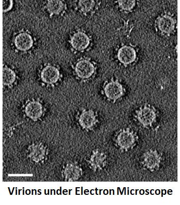
- Observing virus morphology, e.g., influenza or bacteriophages.
- Studying chromatin organization in the nucleus.
Illustrative Example: TEM has been instrumental in virology research, revealing the detailed structure of HIV and SARS-CoV-2 particles. Without TEM, understanding virus assembly and entry into host cells would be nearly impossible.
Scanning Electron Microscope (SEM)
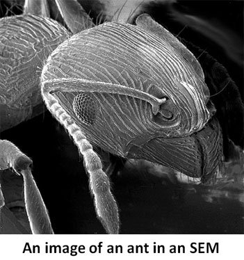
Principle: SEM scans a specimen’s surface with a focused electron beam. Secondary electrons emitted from the surface are detected, creating a three-dimensional image of the specimen’s topography.
Image Characteristics:
- Three-dimensional (3D) surface detail.
- Lower internal resolution compared to TEM.
- Magnification ranges from tens to hundreds of thousands of times.
United States Geological Survey, Public domain, via Wikimedia Commons
Applications:
- Studying the surface structure of pollen grains, insect exoskeletons, or plant leaf surfaces.
- Imaging nanomaterials, polymers, and microfabricated devices.
- Investigating biofilms, microbial colonies, or tissue surfaces.
Illustrative Example: SEM can reveal the spiny surface of a chestnut pollen grain or the detailed morphology of a mosquito’s proboscis—information critical in fields like botany and entomology.
Other Variants
- Scanning Transmission Electron Microscopy (STEM): Combines SEM and TEM capabilities for imaging both surface and internal structures.
- Cryo-Electron Microscopy (Cryo-EM): Specimens are frozen rapidly, preserving their native structure without chemical fixation. Revolutionized structural biology, enabling near-atomic resolution of proteins and complexes.
Illustrative Example: Cryo-EM was used to resolve the structure of the ribosome and coronavirus spike proteins, supporting drug and vaccine development.
J Brew, uploaded on the English-speaking Wikipedia by en:User:Hat’nCoat., CC BY-SA 3.0, via Wikimedia Commons
Specimen Preparation in Electron Microscopy
Proper specimen preparation is critical for achieving clear, artifact-free images in EM. Unlike light microscopy, EM often requires complex and time-consuming preparation.
Key Steps:
Fixation:
- Preserves ultrastructure.
- Chemical fixatives like glutaraldehyde crosslink proteins; osmium tetroxide preserves membranes and adds electron density.
Dehydration and Embedding (for TEM):
- Gradual dehydration using ethanol or acetone.
- Embedded in resin to support ultra-thin sectioning.
Sectioning (for TEM):
- Ultrathin slices (~50–100 nm) cut with an ultramicrotome.
Staining:
- Heavy metal stains like uranyl acetate and lead citrate enhance contrast by scattering electrons.
Surface Preparation (for SEM):
- Critical point drying prevents structural collapse.
- Sputter coating with gold or platinum enhances electron conductivity and image clarity.
Tatiana A. Demina, Hanna M. Oksanen, CC BY 4.0, via Wikimedia Commons
Challenges:
- Time-intensive and technically demanding.
- Risk of artifacts from improper fixation, dehydration, or staining.
- Inability to image living cells directly (except in special environmental EM setups).
Advantages and Limitations
Advantages
- Unmatched resolution: Can reveal structures at nanometer or even atomic scale.
- Ultrastructural detail: Essential for cell biology, virology, and materials science.
- 3D imaging (SEM): Provides surface topography.
- Versatility: Useful for biological, medical, and engineering research.
Limitations
- Expensive: Equipment costs, maintenance, and operation are high.
- Complex specimen prep: Requires expertise; living cells generally cannot be imaged directly.
- Vacuum environment: Limits certain experiments.
- Skill-dependent: Misalignment or poor technique can compromise results.
Applications in Biology and Beyond
Electron microscopy is indispensable across multiple fields:
Cell Biology
- Visualizing mitochondria, lysosomes, Golgi apparatus, and cytoskeletal elements.
- Studying nuclear pores, chromatin condensation, and cell junctions.
Microbiology and Virology
- Imaging bacteria, viruses, flagella, and biofilms.
- Differentiating bacterial morphotypes, such as cocci vs rods, in clinical samples.
Medical Research and Pathology
- Observing ultrastructure of kidney glomeruli or neuromuscular junctions.
- Detecting viral particles in infected tissues (HIV, hepatitis viruses).
- Examining tumor cell morphology at the subcellular level.
Nanotechnology and Materials Science
- Imaging nanoparticles, polymers, semiconductors, and microfabricated devices.
- Surface characterization of materials, revealing defects or irregularities invisible under light microscopy.
Illustrative Example: EM enabled scientists to visualize the structure of SARS-CoV-2, guiding vaccine design and antiviral research. Similarly, TEM reveals the precise arrangement of collagen fibers in connective tissue, crucial for understanding tissue mechanics.
The Future of Electron Microscopy
Electron microscopy continues to evolve with advances in imaging technology, computational analysis, and specimen preservation:
- Cryo-EM: Allows near-atomic resolution of proteins, nucleic acids, and viruses in their native state.
- In-situ EM: Observes dynamic processes in materials and biological samples under controlled conditions.
- Integration with AI and computational reconstruction: Enhances image analysis, segmentation, and modeling of complex structures.
These advances ensure that EM will remain a cornerstone of modern research, bridging the gap between observable cellular processes and molecular-scale phenomena.
Conclusion – Seeing the Invisible World
Electron microscopy has transformed our ability to explore the hidden details of life. By combining high-energy electrons, electromagnetic lenses, and sophisticated detectors, EM surpasses the limits of light microscopy, revealing the ultrastructure of cells, viruses, and materials.
From observing mitochondria in a nerve cell to reconstructing the 3D surface of pollen grains or understanding viral assembly, electron microscopes provide a window into worlds previously considered invisible. For biology students, mastering the principles and applications of EM is not just an academic exercise—it is an essential skill for modern research and discovery.
Electron microscopy reminds us that the microscopic world is vast, intricate, and full of surprises. With patience, skill, and curiosity, you can unlock its secrets and contribute to the next generation of scientific breakthroughs.
Download Quck Note on Electron Microscopy PDF
FAQ on Electron Microscopy
Short Answer Questions (2–3 marks each)
- Define the principle of electron microscopy. (Remembering)
- Differentiate between photons and electrons as illumination sources in microscopy. (Understanding)
- State the typical resolution limit of a light microscope. (Remembering)
- Mention two applications of transmission electron microscopy (TEM) in biology. (Understanding)
- Why is a vacuum environment necessary in electron microscopy? (Understanding)
- List two advantages of cryo-electron microscopy. (Applying)
- What is the function of an electron gun in EM? (Remembering)
- Give one example of how SEM is applied in material science. (Applying)
- Name two heavy metals used for staining in TEM. (Remembering)
- Distinguish between 2D and 3D imaging in TEM and SEM. (Analyzing)
Short Essay Questions (5–7 marks each)
- Explain how the wavelength of electrons improves resolution in EM compared to light microscopy. (Understanding)
- Describe the role of electromagnetic lenses in electron microscopy. (Understanding)
- Compare and contrast TEM and SEM in terms of principles, image characteristics, and applications. (Analyzing)
- Outline the main steps of specimen preparation for TEM. (Remembering/Understanding)
- Discuss the limitations of electron microscopy in studying living cells. (Evaluating)
- Explain the significance of cryo-EM in structural biology. (Applying/Analyzing)
- Illustrate how EM contributed to the structural understanding of SARS-CoV-2. (Applying)
- Analyze the impact of EM in differentiating bacterial morphotypes in clinical samples. (Analyzing)
- Evaluate the role of in-situ EM in modern research. (Evaluating)
- Discuss how artificial intelligence is transforming image analysis in EM. (Evaluating/Creating)
Long Essay Questions (10–15 marks each)
- Describe in detail the principles, types, specimen preparation techniques, and applications of electron microscopy in biology. (Understanding/Analyzing)
- Critically evaluate the advantages and limitations of TEM, SEM, and cryo-EM, giving examples from biology and materials science. (Evaluating)
- Discuss the role of electron microscopy in virology, with reference to HIV and SARS-CoV-2 research. (Analyzing/Evaluating)
- Explain how advances in cryo-EM and AI-based reconstruction are shaping the future of structural biology and nanotechnology. (Evaluating/Creating)
- Propose a research study using electron microscopy to investigate a novel biological or nanomaterial system. Justify your choice of EM type and specimen preparation method. (Creating)
Donload these Questions with Answers PDF
| You may also like... | ||
|---|---|---|
| NOTES | QUESTION BANK | COMPETITIVE EXAMS. |
| PPTs | UNIVERSITY EXAMS | DIFFERENCE BETWEEN.. |
| MCQs | PLUS ONE BIOLOGY | NEWS & JOBS |
| MOCK TESTS | PLUS TWO BIOLOGY | PRACTICAL |