Microscopy is one of the most powerful tools in biology, allowing us to visualize structures invisible to the naked eye. For undergraduate and postgraduate students, understanding microscopy techniques is essential for both coursework and research. Among these methods, dark field microscopy is particularly fascinating because it reveals fine details in transparent and unstained specimens that remain hidden under standard bright field conditions.
Biophysics Notes | Biophysics PPTs | Biophysics MCQs
In this article, we will explore the history, principle, components, sample preparation, applications, advantages, limitations, and modern innovations of dark field microscopy. By the end, you will understand not only how this technique works but also why it remains relevant in today’s biological sciences and beyond.
You may like: MCQ on Dark Field Microscopy
Historical Background
- The idea of using oblique light for microscopy dates back to the 17th century with pioneers like Robert Hooke and Christiaan Huygens.
- However, dark field microscopy became widely practical only in the 19th and early 20th centuries, with the development of specialized dark field condensers.
- Its most famous medical application was in the early diagnosis of syphilis, where the causative bacterium Treponema pallidum could be observed directly from patient samples.
- Even today, dark field microscopy remains an accessible and versatile method in teaching and research labs.
Zephyris at the English-language Wikipedia, CC BY-SA 3.0, via Wikimedia Commons
Principle of Dark Field Microscopy
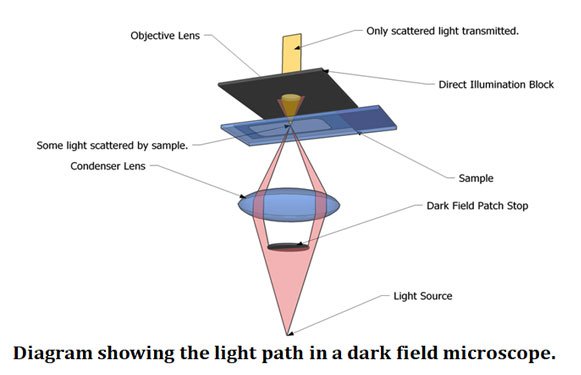
The secret of dark field microscopy lies in scattered light. Instead of passing light directly through a specimen (as in bright field), dark field uses a hollow cone of oblique light created by a special condenser.
- The direct light is blocked.
- Only light scattered by the specimen enters the objective lens.
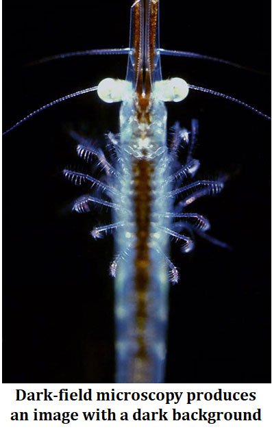
- The specimen appears bright against a dark background.
This method is particularly useful for viewing transparent, thin, or motile samples that lack natural contrast. For example, bacterial flagella, spirochetes, and protozoan cilia become clearly visible.
Key Components of a Dark Field Microscope
- Dark Field Condenser
- Central opaque stop blocks direct light.
- Available in dry and oil immersion versions.
- Objective Lenses
- High numerical aperture (NA) objectives are essential.
- Oil immersion objectives (100x, NA ≥ 1.25) provide maximum resolution.
- Light Source
- Modern systems use halogen or LED light.
- Illumination must be precisely aligned for optimal results.
- Accessories
- Patch stops, phase sliders, and digital cameras enhance versatility.
| You may also like NOTES in... | ||
|---|---|---|
| BOTANY | BIOCHEMISTRY | MOL. BIOLOGY |
| ZOOLOGY | MICROBIOLOGY | BIOSTATISTICS |
| ECOLOGY | IMMUNOLOGY | BIOTECHNOLOGY |
| GENETICS | EMBRYOLOGY | PHYSIOLOGY |
| EVOLUTION | BIOPHYSICS | BIOINFORMATICS |
Sample Preparation for Dark Field Microscopy
- Suitable Specimens: Bacteria, protozoa, blood cells, nanoparticles, thin fibers.
- Wet Mounts: Commonly used for observing living cells and motility.
- Staining: Usually avoided, since dark field already provides contrast.
👉 Teaching Tip: Always keep specimens thin. Thick samples scatter too much light and obscure details.
Applications in Biological Sciences
Microbiology
- Visualization of bacteria, protozoa, and especially spirochetes.
- Essential for studying motility and morphology of living microbes.
Hematology
- Useful in observing red blood cells, platelets, and micro-particles.
Cell Biology
- Ideal for tracking cell movement such as amoeboid locomotion or sperm motility.
Applications in Material and Physical Sciences
- Nanoparticles and Colloids: Dark field reveals scattering colors of nanoparticles, useful in nanoscience.
- Crystals and Fibers: Helps assess microstructures of polymers and films.
- Industrial Quality Control: Detects defects, scratches, or contamination in materials.
Advantages of Dark Field Microscopy
- Provides high contrast without staining.
- Enables real-time observation of living cells.
- Reveals structures invisible in bright field (e.g., flagella, spirochetes).
- Non-invasive and affordable, suitable for teaching labs.
Limitations and Challenges
- Sensitive to sample thickness and preparation.
- Dust or air bubbles may produce artifacts.
- Primarily qualitative, not quantitative.
- Requires precise alignment of condenser and objective.
Comparison with Other Microscopy Techniques
- Bright Field: Needs staining; less useful for transparent cells.
- Phase Contrast: Better for internal structures, but requires specialized optics.
- Fluorescence: Offers molecular specificity, but needs dyes/fluorophores.
👉 Dark field stands out as a simple, label-free technique for highlighting surface and boundary features.
Modern Innovations
Dark field microscopy is evolving with modern tools:
- Digital Imaging: High-resolution cameras for recording and analysis.
- Hybrid Methods: Combining dark field with fluorescence or polarized light.
- Nanoscience: Used to track nanoparticles, exosomes, and biomolecular interactions.
Practical Tips for Students
- Always align the dark field condenser properly.
- Use clean slides and coverslips to avoid artifacts.
- Keep specimens thin for clarity.
- Regularly maintain the microscope for reliable results.
Quick Guide to Dark Field Microscopy
🔬 What is Dark Field Microscopy?
- Optical microscopy technique using oblique light.
- Only scattered light forms the image → specimen appears bright on a dark background.
- Best for transparent, unstained, and motile samples.
⚙️ Key Components
- Dark Field Condenser – blocks central light, allows oblique rays.
- High NA Objectives – especially oil immersion (100x, NA ≥ 1.25).
- Light Source – LED or halogen; needs precise alignment.
- Accessories – patch stops, phase sliders, digital cameras.
🧫 Sample Preparation
- Works best with thin, clean specimens.
- Wet mounts for live cells and motility studies.
- Avoid staining (adds unwanted scattering).
🧬 Biological Applications
- Detects bacteria, protozoa, spirochetes (e.g., Treponema).
- Observes blood cells, sperm motility, amoeboid movement.
- Ideal for cell boundaries and flagella.
✅ Advantages
- High contrast without staining.
- Real-time imaging of living cells.
- Reveals structures invisible in bright field.
⚠️ Limitations
- Sensitive to sample thickness.
- Artifacts from dust or air bubbles.
- Qualitative, not quantitative.
- Requires careful alignment.
🔄 Comparison
- Bright Field: Needs staining; less effective for transparent cells.
- Phase Contrast: Reveals internal details.
- Fluorescence: Molecular specificity with dyes.
- Dark Field: Best for surface structures & motility.
Conclusion and Future Perspectives
Dark field microscopy remains one of the most student-friendly yet research-relevant techniques. For biology undergraduates and postgraduates, it provides a practical way to study living cells, microorganisms, and microstructures without complicated staining or expensive equipment.
As microscopy continues to integrate with digital tools and nanotechnology, dark field imaging will likely find new applications in biomedicine, environmental monitoring, and material sciences. For students, mastering this technique builds a strong foundation for exploring more advanced microscopy methods.
References (Suggested for Further Reading)
- Bradbury, S., & Bracegirdle, B. (1998). Introduction to Light Microscopy. BIOS Scientific Publishers.
- Murphy, D. B., & Davidson, M. W. (2012). Fundamentals of Light Microscopy and Electronic Imaging (2nd ed.). Wiley-Blackwell.
- Porter, K. R. (1973). “Dark-field microscopy and its application to microbiology.” Annual Review of Microbiology, 27, 1–24.
- Unger, A., & Beck, T. (2011). “Dark-field microscopy in nanoscience.” Analytical and Bioanalytical Chemistry, 399, 2717–2729.
- LeGresley, P. A., & Cooke, D. J. (2020). “Applications of dark-field microscopy in biomedical sciences.” Journal of Microscopy, 280(3), 135–150.
| You may also like... | ||
|---|---|---|
| NOTES | QUESTION BANK | COMPETITIVE EXAMS. |
| PPTs | UNIVERSITY EXAMS | DIFFERENCE BETWEEN.. |
| MCQs | PLUS ONE BIOLOGY | NEWS & JOBS |
| MOCK TESTS | PLUS TWO BIOLOGY | PRACTICAL |