What is Chromosome Banding?
Chromosome banding is an essential technique used in chromosome karyotyping to identify normal and abnormal chromosomes for clinical and research purposes. This technique involves staining the chromosomes to create distinctive patterns or bands on the chromosomes. The unique banding pattens helps to identify individual chromosomes and to analyse the structural abnormalities. The ability to visualize chromosomal bands has led to significant advancements in various areas of genetics, including diagnosis of genetic disorders, evolutionary studies, and cancer research. In this article, we will discuss the principles behind chromosome banding techniques and delve into the different types of banding methods employed in cytogenetics.
Principles of Chromosome Banding Technique
Ø Chromosome banding techniques exploit variations in DNA content, base composition, and chromatin condensation of a chromosome to create unique patterns of bands or stripes.
Ø These bands serve as distinctive landmarks that facilitate the identification and analysis of chromosomes.
Ø Process of chromosome banding typically involves THREE steps:
(1). Cell Culturing and Preparation
Ø Cells from blood, bone marrow, or tissue samples are cultured to promote their division.
Ø This ensures the availability of actively dividing cells for the analysis.
Ø Once a suitable number of cells are obtained, they are treated with a mitotic spindle inhibitor (e.g., colchicine) to arrest them in the metaphase.
Ø At metaphase, the chromosomes will be maximum condensed and visible.
You may also like: Cell Biology Notes | Cell Biology PPT | Cell Biology MCQ |
(2). Chromosome Staining
Ø The arrested cells are then treated with a suitable staining solution.
Ø The stain interacts with the chromosomal DNA, causing distinct regions of the chromosomes to take up stain at different intensities.
Ø This staining results in the formation of alternating light and dark bands along the length of the chromosomes.
(3). Microscopic Analysis
Ø The stained chromosomes are examined under a light microscope equipped with appropriate imaging techniques to enhance the visibility of the bands.
Ø The resulting pattern of bands is unique to each chromosome and provides valuable information about their structure and arrangement.
| You may also like NOTES in... | ||
|---|---|---|
| BOTANY | BIOCHEMISTRY | MOL. BIOLOGY |
| ZOOLOGY | MICROBIOLOGY | BIOSTATISTICS |
| ECOLOGY | IMMUNOLOGY | BIOTECHNOLOGY |
| GENETICS | EMBRYOLOGY | PHYSIOLOGY |
| EVOLUTION | BIOPHYSICS | BIOINFORMATICS |
Chromosome Banding Techniques
Ø Several chromosome banding techniques have been developed
Ø Each method yields specific patterns of bands that aid in chromosome identification and analysis.
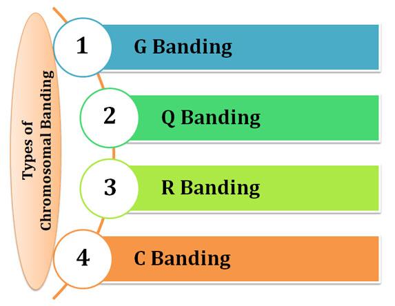
Ø Some of the most widely used chromosome banding methods are:
(a). G Banding (Giemsa)
(b). Q Banding (Quinacrine)
(c). R Banding (Reverse)
(d). C Banding (Constitutive Heterochromatin)
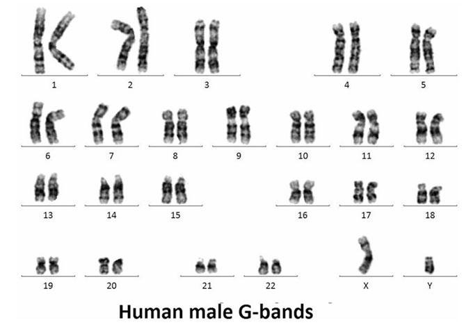
(a). Giemsa (G) Banding
Ø Banding chromosome with Giemsa staining is called G-banding.
Ø Giemsa solution is a mixture of methylene blue, eosin, and Azure.
Ø G banding is one of the most used chromosome banding techniques and it stains the heterochromatic regions of the chromosomes.
Ø When chromosomes are treated with Giemsa dye, it produces characteristic dark and light bands along the chromosomes.
Ø Giemsa is specific for the phosphate groups of DNA and attaches itself to regions of DNA where there are high amounts of adenine-thymine bonding.
Ø G-banding is valuable for routine cytogenetic analysis, karyotyping, and identifying structural abnormalities of the chromosome.
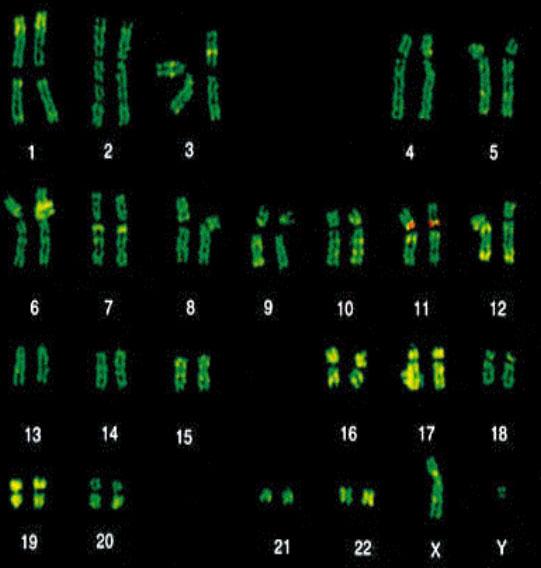
(b). Quinacrine (Q) Banding
Ø Banding of chromosome with Quinacrine mustard is called Q banding.
Ø Quinacrine is a yellow fluorescent dye is used to stain chromosomes.
Ø Under UV light, the chromosomes exhibit fluorescent bands that can be observed and analysed.
Ø Q banding create pattern similar to G banding (stain heterochromatin).
Ø Most part of the stained DNA in Q banding is heterochromatin.
Ø Quinacrine binds both regions rich in AT and in GC, but only the AT-quinacrine-complex fluoresces.
Ø Since regions rich in AT are more common in heterochromatin than in euchromatin, these regions are labelled preferentially.
Ø Q-banding is particularly useful for identifying chromosomes with high AT-rich regions.
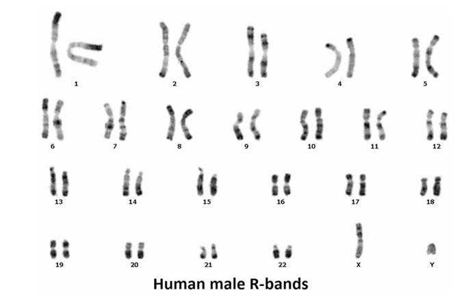
(c). Reverse (R) Banding
Ø Reverse banding is a reverse variant of G-banding
Ø Here the chromosomes are treated with a mild heat shock before staining with Giemsa.
Ø This results in the reversal of the typical G-banding pattern, with dark bands becoming light and vice versa.
Ø R-banding can provide complementary information to G-banding and aid in the identification of specific chromosomal regions.
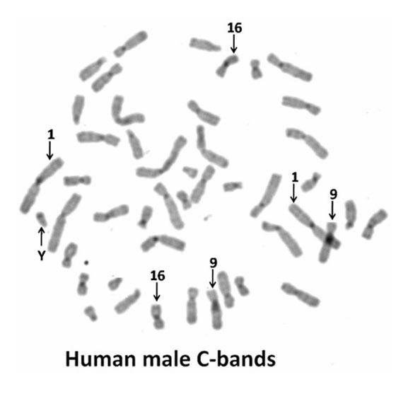
(d). C-Banding
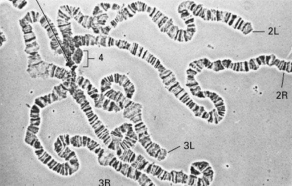
Ø It is the constitutive heterochromatin banding. Also called Centromere banding.
Learn more: Classification of Chromosome PPT
Ø C banding specifically stains the centromeric regions of chromosomes.
Ø It reveals the distribution of heterochromatic regions, which are rich in repetitive DNA sequences.
Ø C-banding is useful for identifying centromeres and studying chromosomal rearrangements
Significance and Applications of Chromosome Banding
Chromosome banding techniques have applications across various fields of genetics and biology.
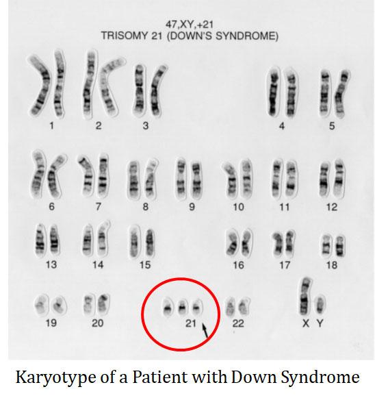
(a). Karyotyping in Clinical Diagnostics
Ø These techniques are crucial for diagnosing genetic disorders, such as Down syndrome (Trisomy 21) and Turner syndrome (Monosomy X), by identifying numerical and structural chromosomal abnormalities.
(2). Cancer Research
Ø Chromosome banding helps identify chromosomal aberrations in cancer cells, aiding in cancer diagnosis, prognosis, and treatment planning.
Learn more: Philadelphia Chromosome and Cancer
(3). Evolutionary and Comparative Genomic Studies
Ø Chromosome banding can be used to compare the chromosomal organization and evolution of different species.
Ø It helps in studying genome similarities, differences, and evolutionary relationships among species.
(4). Genome Mapping
Ø Chromosome banding aids in constructing detailed karyotypes and mapping specific genes or loci to specific chromosomal regions.
(5). Forensic Investigations
Ø Chromosome banding techniques can assist in forensic DNA analysis, particularly in cases where chromosomal abnormalities are involved.
(6). Genetic Counselling
Ø Chromosome banding results can be used by genetic counsellors to provide information and guidance to individuals and families affected by chromosomal disorders.
Ø This helps in understanding the inheritance patterns, recurrence risks, and potential implications for future generations.
Summary: Chromosome banding techniques have become indispensable tools in modern cytogenetics, enabling researchers to unravel the complex genomic landscape within cells. The distinctive patterns of bands produced by these techniques provide insights into chromosome structure, organization, and function, leading to advancements in diagnostics, cancer research, evolutionary studies, and more.
<<< Back to CYTOLOGY Notes Page
| You may also like NOTES in... | ||
|---|---|---|
| BOTANY | BIOCHEMISTRY | MOL. BIOLOGY |
| ZOOLOGY | MICROBIOLOGY | BIOSTATISTICS |
| ECOLOGY | IMMUNOLOGY | BIOTECHNOLOGY |
| GENETICS | EMBRYOLOGY | PHYSIOLOGY |
| EVOLUTION | BIOPHYSICS | BIOINFORMATICS |
| You may also like... | ||
|---|---|---|
| NOTES | QUESTION BANK | COMPETITIVE EXAMS. |
| PPTs | UNIVERSITY EXAMS | DIFFERENCE BETWEEN.. |
| MCQs | PLUS ONE BIOLOGY | NEWS & JOBS |
| MOCK TESTS | PLUS TWO BIOLOGY | PRACTICAL |