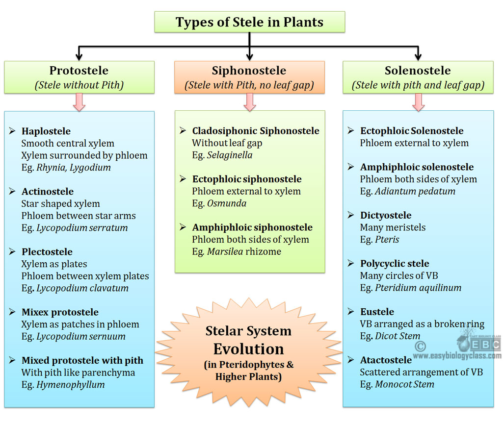
Stelar Evolution in Pteridophytes
What is stele? What are the components of stele?
Ø Stele is the central cylinder or core of vascular tissue in higher plants.
Ø The stele consists of xylem, phloem, pericycle and medullary rays and pith if present.
Ø The term ‘stele’ was for the first time used by Van Tieghem and Douliot in 1886 in their ‘Stelar Theory’.
What is ‘stellar’ theory’?
Ø Proposed by Van Tieghem and Douliot in 1886.
Ø Major highlights in stellar theory are:
$. The stele is a real entity and present universally in all axis of higher plants.
$. The primary components of stele are xylem and phloem.
$. Tissues like pericycle, medullary rays and pith are also the components of stele.
$. ‘Stelar theory’ also says that the cortex and the stele are the two fundamental parts of a shoot system.
$. Both these components (stele and cortex) are separated by the endodermis.
$. In higher vascular plants (Pteridophytes, Gymnosperms and Angiosperms), the leaf traces are large, and it appears that they play an important role in the vascular system of the axis.
$. The whole set-up of leaf traces appears as a composite structure in these plants.
$. Such composite structures do not remain within the limits of stellar theory of Van Tieghem and Douliot.
Different types of steles in plants (Pteridophytes and higher plants)
Ø On the basis of ontogeny and phylogeney, there are THREE broad categories of steles in vascular plants.
Ø They are:
(1). Protostele
(2). Siphonostele
(3). Solenostele
Ø Some authors recognize only two categories (Protostele and Siphonostele). They consider Solenostele as a sub-category of Siphonostele.
(1). Protostele
Ø A stele in which the vascular core consists of a solid core of xylem and it is surrounded by phloem, pericycle and endodermis is called protostele.
Ø Pith is absent in protostele.
Ø Protostele represents the simplest stellar organization in vascular plants.
Ø Majority of the Pteridophytes show protostelic condition in their rhizome, stem or roots.
Ø Protostele is considered as the most primitive stellar organization in plants.
Ø There are FIVE types of protosteles in Pteridophytes, they are: (a) Haplostele, (b) Actinostele, (c) Plectostele, (d) Mixed protostele and (e) Mixed protostele with pith.
(a). Haplostele
Ø A protostele with a smooth core of xylem surrounded by uniform layers of phloem.
Ø Named by Brebner in 1902.
Ø Considered as the most primitive type of protostele.
Ø Usually present in fossil genera like Rhynia and Horneophyton
Ø Example: Selaginella, Gleichenia and Lygodium.
 (b). Actinostele
Ø Protostele with xylem core having radial ribs or arms.
Ø Xylem is star shaped or stellate, hence the name.
Ø The phloem is NOT present in a continuous manner.
Ø Phloem occurs as separate patches between the arms of xylem.
Ø Named by Brebner in 1902.
Ø Example: Asteroxylon, Psilotum, Lycopodium serratum.

(c). Plectostele
Ø Xylem occurs as several plates which are more or less parallel to each other.
Ø Such xylem plates are alternated with phloem patches.
Ø Named by Zimmermann in 1930.
Ø Example: Lycopodium clavatum.
 (d). Mixed protostele
Ø Xylem is divided into several units or groups.
Ø Each xylem units are scatteredly arranged inside the ground mass of phloem.
Ø Example: Lycopodium cernuum.
| You may also like NOTES in... | ||
|---|---|---|
| BOTANY | BIOCHEMISTRY | MOL. BIOLOGY |
| ZOOLOGY | MICROBIOLOGY | BIOSTATISTICS |
| ECOLOGY | IMMUNOLOGY | BIOTECHNOLOGY |
| GENETICS | EMBRYOLOGY | PHYSIOLOGY |
| EVOLUTION | BIOPHYSICS | BIOINFORMATICS |
(d). Mixed protostele with pith
Ø Advanced type of stele among protosteles.
Ø The formation of pith in the stele started here for the first time in the evolution.
Ø Stele is similar to mixed protostele.
Ø Here small patches of parenchymatous region occur in association xylem strands.
Ø Mixed protostele with pith is considered as a connecting link between protostele and siphonostele.
Ø Example: Hymenophyllum demissum, Lepidodendron selaginoides.

(2). Siphonostele
Ø A stele with pith (medulla) at the centre is called siphonostele.
Ø The central core of pith is surrounded by the xylem.
Ø Siphonostele is an advanced type of stele than protostele.
Origin of siphonostele
Ø The siphonostele is derived from protostele by the formation of pith in the centre.
Ø The centrally placed xylem core in a protostele is replaced by parenchymatous pith.
Ø Different stages of changing of protostele to siphonostele can be observed in the transverse sections at different levels in Gleichenia, Osmunda and Anemia.
Ø There are TWO views regarding the origin of pith in the siphonostele:
(i). Inter-stelar origin of pith:
$. According to this theory, the innermost (centrally placed) vascular tissue in a protostele changes into parenchymatous cells.
$. This theory is proposed by Bower in 1923 and supported by Fahn in 1960.
$. This theory is the most widely accepted theory of pith development.
(ii). Extra-stelar origin of pith:
$. According to this theory, the pith is formed as a result of the invasion of cortical parenchymatous cells into the stele.
$. The invasion of pith occurs through the leaf gap or branch gap.
$. Thus pith and cortex are homogenous structures according to this theory.
$. This theory is proposed by Jeffery.
$. It is not accepted by most of the authors since in many Pteridophytes there is stele without leaf gaps but having pith in the centre.
Different types of siphonosteles
Ø Based on the position and distribution of phloem, TWO types of siphonostles reported among Pteridophytes are (a) Ectophloic siphonostele and (b) Amphiphloic siphonostele.
(a). Ectophloic siphonostele
Ø Phloem is present only on the external side of the xylem.
Ø The pith is at the central position.
Ø Phloem is externally surrounded by pericycle and endodermis.
Ø Leaf traces present, but leaf gap absent
Ø Example: Osmunda, Schizaea
(b). Amphiphloic siphonostele
Ø Phloem is present on both sides of the xylem (external and internal phloem).
Ø The central portion of the stele is occupied by pith.
Ø Xylem on its inner side is surrounded by inner phloem, inner pericycle and inner endodermis.
Ø Xylem on its outer side is surrounded by outer phloem, outer pericycle and outer endodermis.
Ø Example: Marsilea, Adiantum

(3). Solenostele
Ø Solenostele is actually a sub category of siphonostele.
Ø A siphonostele which is perforated at the place of origin of leaf trace is called solenostele.
Ø In simple term, siphonostele with leaf gap is called solenostele.
Different types of Solenostele
Ø Six different types of solenostele can be observed among plant kingdom.
Ø They are (a) Ectophloic solenostele, (b). Amphiphloic solenostele, (c) Dictyostele, (d) Eustele and (e) Atactostele.
(a). Ectophloic solenostele
Ø Derived from ectophloic siphonostele.
Ø Thus phloem is present only on the outer side of the xylem.
(b). Amphiphloic solenostele
Ø Derived from amphiphloic siphonostele.
Ø Thus phloem is present on both sides of the xylem.
Ø Phloem in both sides is intern surrounded by pericycle and endodermis.
Ø Example: Adiantum pedatum

(c). Dictyostele
Ø Solenostele that is broken into a network of separate vascular strands are called dictyostele.
Ø This breaking-up of stelar core is due to the presence of large number of leaf gaps.
Ø Each such separate vascular strand is called meristele.
Ø Example: Pteris, Adiantum capillus-veneris.
(d). Poly-cyclic stele
Ø Here the stele is present as two or more concentric cylinders.
Ø Poly-cyclic stele will be always solenostelic in nature.
Ø Poly-cyclic stele may be polycyclic solenostele or polycyclic dictyostele

(d). Eustele
Ø If the stele is split into distinct collateral vascular bundles, then it is called eustele.
Ø It is modified ectophloic siphonostele.
Ø Spitting of the original stelar core takes place due to the overlapping of large number of leaf gaps.
Ø Individual vascular bundles in the eustele are arranged as broken ring in the ground tissue.
Ø Example: dicot stem primary structure

(e). Atactostele
Ø Similar to eustele.
Ø Both the individual vascular bundles are scatteredly distributed in the ground tissue
Ø Example: Monocot stem

Test your understanding…
- What is stelar theory?
- Define ‘stele’.
- Who proposed the ‘stelar theory’?
- What are the main points in ‘stelar theory’?
- Name the three major categories of steles in vascular plants.
- What is meant by protostele?
- Describe different types of Protostels with examples.
- Differentiate actinostele and haplostele.
- What is meant by siphonostele?
- Describe different types of siphonosteles with examples.
- What is meant by solenostele?
- Describe different types of solenosteles with examples?
- Define meristele.
- What is atactostele?
- What is meant by Eustele?
- Describe dictyostele with example.
- Differentiate ectophloic and amphiphloic solenostele.
- Write an essay on stelar evolution in Pteridophytes with suitable examples and neat diagrams.
| You may also like... | ||
|---|---|---|
| NOTES | QUESTION BANK | COMPETITIVE EXAMS. |
| PPTs | UNIVERSITY EXAMS | DIFFERENCE BETWEEN.. |
| MCQs | PLUS ONE BIOLOGY | NEWS & JOBS |
| MOCK TESTS | PLUS TWO BIOLOGY | PRACTICAL |


 (b). Actinostele
 (d). Mixed protostele
Like this subject
Lectures are easy and very helpful .