Bacterial Staining Techniques
Why Is Bacterial Staining Necessary?
Unstained bacterial cells are transparent and difficult to distinguish from the surrounding medium. Staining increases contrast and makes microorganisms clearly visible under a microscope.
- Increases contrast between cells and background
- Enhances visualization of morphology (shape and arrangement)
- Preserves cell structure for examination
- Differentiates bacteria into groups
Before staining, however, cells must first undergo fixation.

Fixation: The First Step in Bacterial Staining
Fixation is the process of preserving cells and attaching them firmly to the microscope slide. The goal is to maintain cellular structures as close as possible to their natural state while preventing enzymatic degradation.
Fixation performs three major functions:
- Kills microorganisms
- Preserves internal and external structures
- Prevents cells from being washed off during staining
1. Heat Fixation
In this method, an air-dried bacterial smear is gently passed through a flame. Heat fixation preserves overall cell morphology but does not maintain fine internal structures.
2. Chemical Fixation
Chemical fixatives penetrate cells and stabilize proteins and lipids. Common chemical fixatives include ethanol, formaldehyde, glutaraldehyde, and acetic acid. This method is especially useful for delicate or larger microorganisms.
Understanding Dyes in Microbiology
Dyes used in microbiology share two key characteristics:
- They contain chromophores — chemical groups responsible for color.
- They bind to cells through ionic, covalent, or hydrophobic interactions.
Since bacterial cell surfaces are typically negatively charged, positively charged dyes bind readily to them.
Types of Dyes
Basic Dyes
Examples: methylene blue, crystal violet, safranin, malachite green.
- Positively charged
- Bind to negatively charged bacterial cells
- Most commonly used in bacteriology
Acid Dyes
Examples: eosin, rose bengal, acid fuchsin.
- Negatively charged
- Bind to positively charged cellular structures
Effect of pH on Staining
The pH of the staining environment affects dye binding because cellular charge varies with pH. Acid dyes work best in acidic conditions, while basic dyes are more effective in alkaline environments.
Some dyes bind through special mechanisms:
- The Feulgen method stains DNA via covalent bonding.
- Sudan Black stains lipids because it is lipid-soluble.
Simple Staining Technique
Simple staining involves the use of a single dye. It is quick, easy, and widely used to determine:
- Size
- Shape
- Arrangement of bacteria
The procedure includes applying the stain, washing off excess dye, and drying the slide. Basic dyes such as crystal violet or methylene blue are commonly used.
While simple staining reveals morphology, it does not differentiate between bacterial types.
Differential Staining in Microbiology
Differential staining distinguishes bacteria into groups based on structural differences.
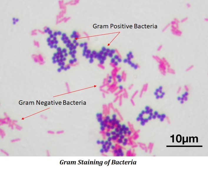
Gram Staining
The Gram stain, developed in 1884 by Christian Gram, is the most important staining technique in microbiology. It divides bacteria into:
- Gram-positive bacteria
- Gram-negative bacteria
Steps of Gram Staining
- Crystal violet (primary stain)
- Iodine (mordant)
- Alcohol or acetone (decolorizer)
- Safranin (counterstain)
Gram-positive bacteria retain crystal violet and appear purple. Gram-negative bacteria lose the primary stain and appear pink after counterstaining. This difference is due to variations in cell wall structure.
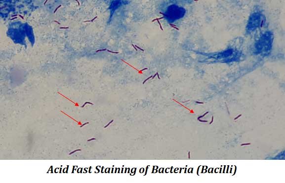
Acid-Fast Staining
Some bacteria, particularly species of Mycobacterium, resist conventional staining because of their waxy cell walls rich in mycolic acid.
The Ziehl-Neelsen method is used for acid-fast staining:
- Cells are heated with basic fuchsin and phenol.
- Acid-alcohol is used for decolorization.
- Methylene blue is applied as a counterstain.
Acid-fast bacteria remain red, while non-acid-fast bacteria appear blue.
This technique is essential for identifying pathogens such as Mycobacterium tuberculosis and Mycobacterium leprae.
Staining Specific Bacterial Structures
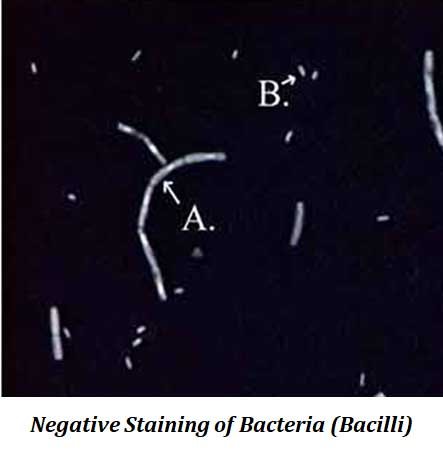
1. Negative Staining (Capsule Staining)
In negative staining, bacteria are mixed with India ink or Nigrosin. The dye stains the background, not the cell.
Bacteria appear as light bodies against a dark background. Capsules appear as clear halos surrounding the cell.
This method causes minimal distortion of cell shape.
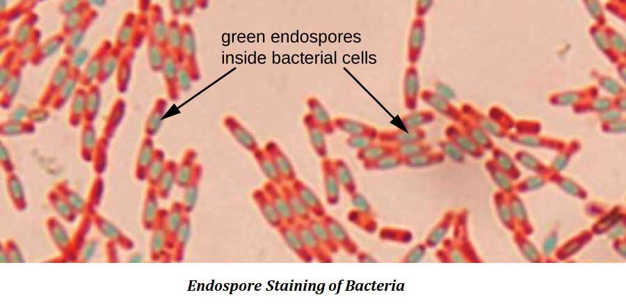
2. Endospore Staining
Some bacteria, such as Bacillus and Clostridium, form resistant endospores.
The Schaeffer-Fulton method is commonly used:
- Malachite green is applied with heat.
- The slide is washed with water.
- Safranin is used as a counterstain.
Endospores appear green, while vegetative cells appear pink or red.
3. Flagella Staining
Flagella are extremely thin structures used for locomotion and are not normally visible under a light microscope.
- Mordants increase their thickness.
- Special stains like pararosaniline or basic fuchsin are applied.
Flagella staining helps determine the presence and arrangement of flagella, which is important for bacterial identification.
Conclusion
Bacterial staining techniques are cornerstone methods in microbiology. They transform invisible, transparent cells into clearly distinguishable structures, allowing scientists to study morphology, classify bacteria, and identify pathogens.
From simple staining for morphology to Gram staining and acid-fast staining for classification, and specialized techniques for capsules, endospores, and flagella — each method provides valuable diagnostic and taxonomic information.
A strong understanding of bacterial staining principles is essential for laboratory practice, academic study, and competitive examinations in microbiology and life sciences.
| You may also like NOTES in... | ||
|---|---|---|
| BOTANY | BIOCHEMISTRY | MOL. BIOLOGY |
| ZOOLOGY | MICROBIOLOGY | BIOSTATISTICS |
| ECOLOGY | IMMUNOLOGY | BIOTECHNOLOGY |
| GENETICS | EMBRYOLOGY | PHYSIOLOGY |
| EVOLUTION | BIOPHYSICS | BIOINFORMATICS |
Study Offline!! Download this Note in PDF
🧪 Test Your Knowledge: Bacterial Staining MCQs with Answers
1. Why is bacterial staining necessary?
A. To make bacteria grow faster
B. To increase contrast and make bacteria visible
C. To kill all microorganisms permanently
D. To change bacterial shape
2. What is the main purpose of fixation in bacterial staining?
A. To color the bacteria
B. To enlarge the bacteria
C. To preserve cells and attach them to the slide
D. To remove all cell structures
3. Which of the following is a chemical fixative?
A. Crystal violet
B. Ethanol
C. Safranin
D. Methylene blue
4. Basic dyes are typically:
A. Negatively charged
B. Positively charged
C. Neutral in charge
D. Colorless
5. Which staining technique uses only one dye?
A. Gram staining
B. Acid-fast staining
C. Simple staining
D. Endospore staining
6. In Gram staining, what is the role of iodine?
A. Decolorizer
B. Counterstain
C. Mordant
D. Primary stain
7. After Gram staining, Gram-negative bacteria appear:
A. Purple
B. Green
C. Pink
D. Blue
8. Acid-fast bacteria resist staining due to:
A. Thick peptidoglycan layer
B. Presence of capsules
C. Waxy cell wall rich in mycolic acid
D. Presence of flagella
9. In negative staining, what gets stained?
A. The bacterial cell
B. The capsule only
C. The background
D. The nucleus
10. In the Schaeffer-Fulton method, endospores appear:
A. Pink
B. Blue
C. Green
D. Purple
<<< Back to Microbiology Notes Page
| You may also like... | ||
|---|---|---|
| NOTES | QUESTION BANK | COMPETITIVE EXAMS. |
| PPTs | UNIVERSITY EXAMS | DIFFERENCE BETWEEN.. |
| MCQs | PLUS ONE BIOLOGY | NEWS & JOBS |
| MOCK TESTS | PLUS TWO BIOLOGY | PRACTICAL |